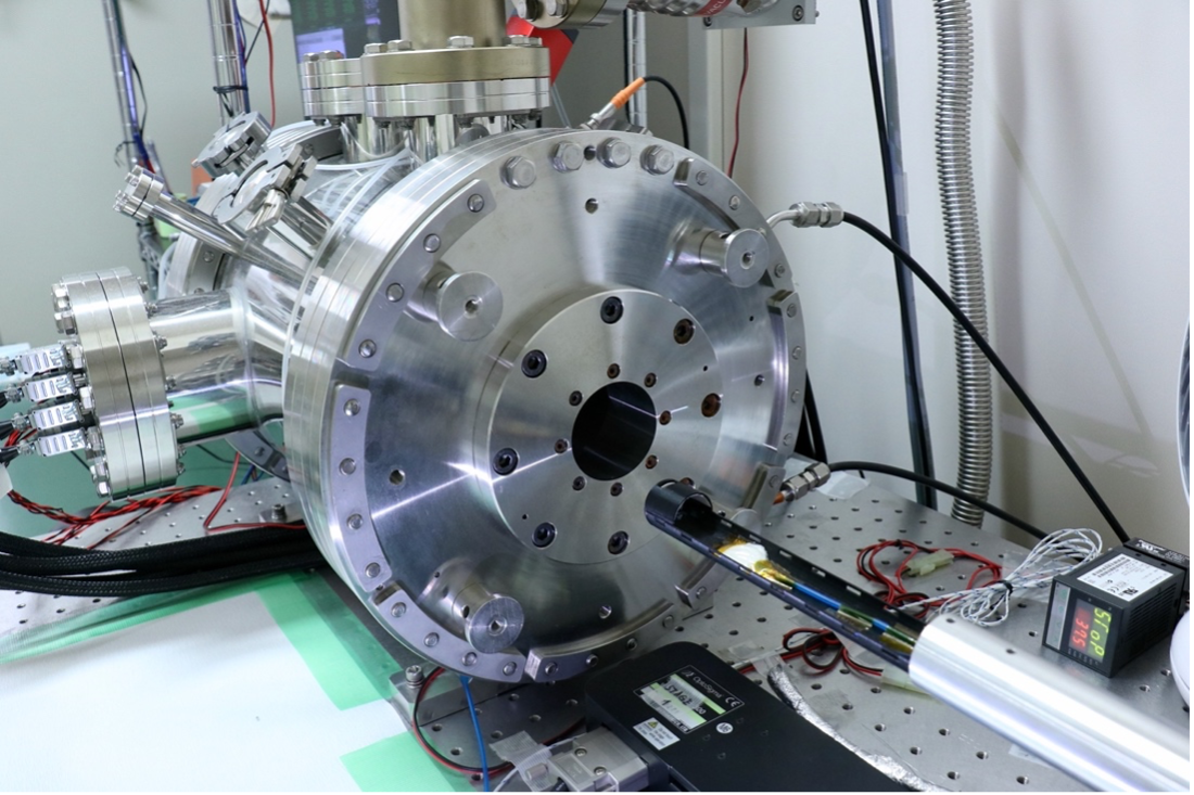
The ultra-high resolution small animal SPECT system used in this study, developed primarily by researchers at Kavli IPMU.
Researchers have shown it is possible to image small animal tissue clearly to several hundred micrometers using multi-probe imaging, reports a recent study in Scientific Reports.
This technique could be useful in various fields of medical research because it enables researchers to observe the microstructure of small animal tissues, and clarify the localization and interaction of multiple molecules such as microscopic metastatic lesions of cancer cells.
Single-photon emission tomography (SPECT) is currently used for molecular imaging in both animals and humans. However, the technology faces several limitations, including relatively low spatial resolution and challenges associated with the simultaneous use of multiple probes.
A team of researchers, led by Kavli Institute for the Physics and Mathematics of the Universe (Kavli IPMU) Project Assistant Professors and National Cancer Center Center for Advanced Biomedical Research and Development (NCCER) Visiting Researcher Atsushi Yagishita and Shin’ichiro Takeda, and involving researchers from Kavli IPMU, NCCER, and Keio University, resolved these problems using a SPECT system equipped with a cadmium telluride (CdTe) semiconductor detector that was previously used for space observations.
This device was initiated in development by High Energy Accelerator Research Organization Professor Emeritus Hirotaka Sugawara, Kavli IPMU’s Specially Appointed Assistant Professor Shin’ichiro Takeda and Tadashi Orita, and others during their tenure at the Okinawa Institute of Science and Technology Graduate University (OIST). There, by applying the spectral analysis methods used in the analysis of astronomical observation data, they succeeded in obtaining high spatial resolution images for each of the multiple radioactive nuclide probes used simultaneously (Takeda et al., IEEE TRPMS 2023).
Using the device, the researchers this time performed SPECT imaging of submillimeter zeolite spheres absorbed with 125I- and subsequently imaged 125I-accumulated spheroids, cells that aggregates to form a sphere-like shape, which were 200–400 μm in size within an hour. They successfully captured clear and quantitative images. Furthermore, their dual-radionuclide phantom imaging revealed a distinct image of the submillimeter sphere absorbed with 125I- immersed in a 99mTc-pertechnetate solution, and provided a fair quantification of each radionuclide.
Then, the team performed in vivo imaging on a cancer-bearing mouse with lymph node micro-metastasis using dual-tracers. The results displayed dual-tracer images of lymph tract by 99mTc-phytic acid and the submillimeter metastatic lesion by 125I-, shown to align with the immunofluorescence image (Figure 2).
The researchers say their method could provide benefits to biological research, pharmaceutical research, and medical research.
Details of their study were published in Scientific Reports on 9 November.
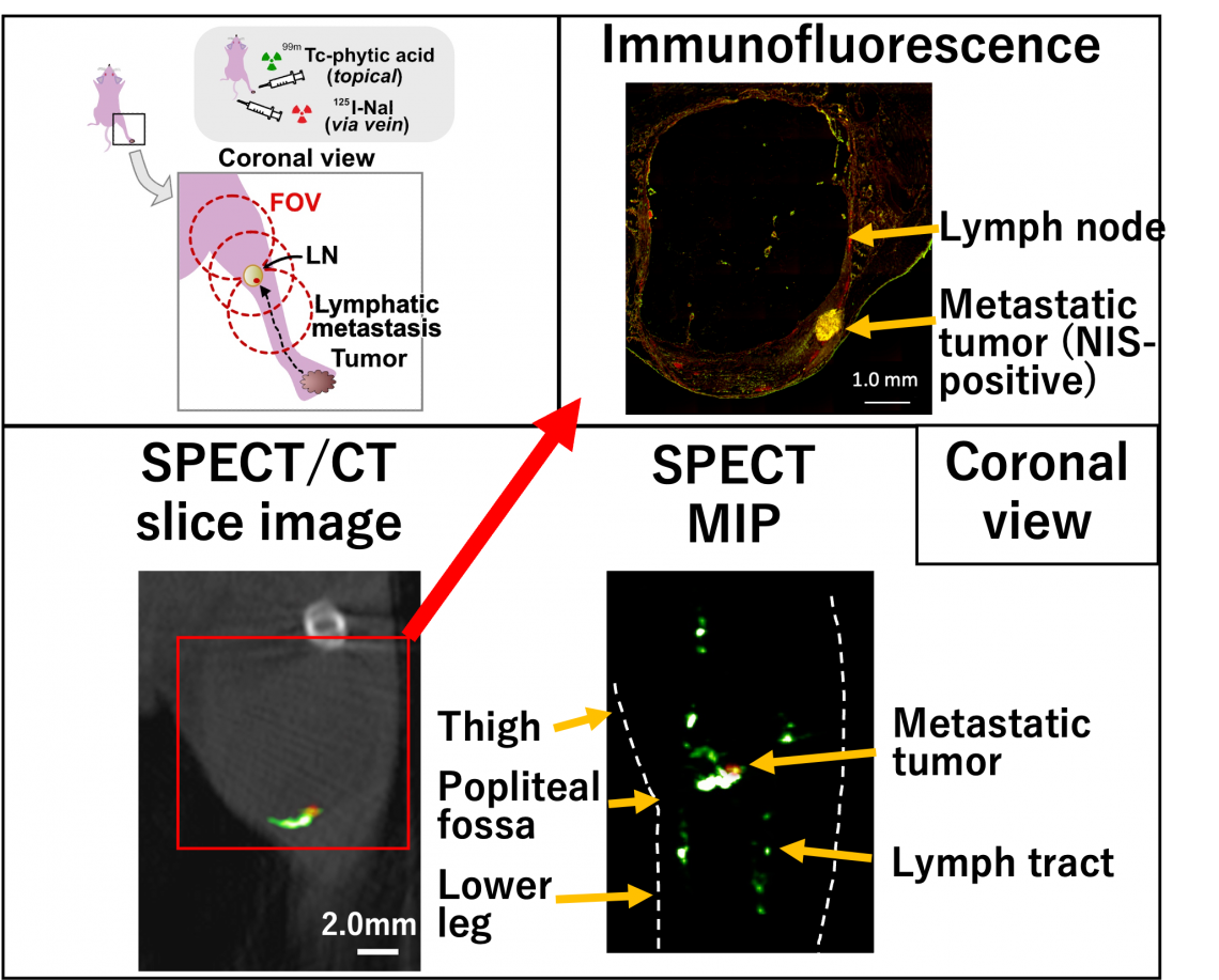
A schematic diagram of the animal experiments conducted in this study. (Upper left box) SPECT imaging was performed four weeks after 4T1-mNIS cancer cells were implanted in the foot pad. The red dashed circles indicate the field of view for SPECT. The tracer for lymphatic channels and nodes, 99mTc-phytate, was topically administered in the foot pad, and the tumor tracer 125I -NaI was administered intravenously. (Bottom box) The left image shows a SPECT/CT image of the lymph node and its surrounding. Photons from 125I (25-30 keV) are colored in red and those from 99mTc (138-142 keV) are colored in green. The right image shows a SPECT projection image. The green tubular structure by 99mTc-phytate indicates lymphatic vessels, and the small red (125I -NaI) spots on the lymphatic vessels indicates the metastatic tumors.
(Upper right box) An immunofluorescence image shows a NIS-positive (yellow) metastatic tumor less than 1 mm in size within the lymph node. This location is consistent with the location of the tumor shown in the SPECT/CT image in the lower left image.